• Contact us
  • Young GI
  • Patients
  • Industry
  • Press
Create myUEG account
  • About
    • To landing page
      • Who we are
      • Boards & Committees
      • Governance
      • Secretariat
      • What we do
      • Strategic Drivers
      • Annual Report
      • Our Members
      • Specialist Member Societies
      • National Member Societies
      • Community
      • myUEG
      • Women in GI
      • Young GIs
  • Week
    • To landing page
      • Register
      • Fees
      • CME Accreditation
      • Science
      • Abstracts
      • Programme
      • Postgraduate Teaching
      • Networking
      • Industry Programme
      • Acknowledgements
      •  
      • Congress Resources
      • Week 24/7 recordings
  • Education
    • To landing page
      • Online Education
      • Online Courses
      • Webinars
      • Virtual Summer School
      • Face-to-face Education
      • Masterclass
      • Clinical Visiting Fellowships
      • Education Resources
      • Mistakes in...
      • Articles
      • Image Hub
      • CME in Europe
  • Research
    • To landing page
      • Support
      • Research Prize
      • Research Fellowship
      • Researchers Network
      • Research Webinars
      • Advocacy
      • Position Papers
      • White Book
  • Quality of Care
    • To landing page
      • Search Guidelines
      • Standards & Guidelines Repository
      • Non-English Guidelines
      •  
      • GI Guidelines App
      • Develop & Implement
      • Essentials for a good guideline
      • Get Funding
      • Guideline Webinars
      • Projects
      • Quality of Care Initiatives
      • Quality Standards
  • Public Affairs
    • To landing page
      • Advocacy
      • Manifesto on Digestive Health
      • Core Positions
      • Position Papers
      • Stakeholder Collaboration
      • MEP Digestive Health Group
      • Key Topics
      • Our Supporters
      • #EUNewsline →
  • Publications
    • To landing page
      • Publications
      • Congress Publications
      • Research Publications
      • Public Affairs Publications
      • Education Publications
      • UEG Publications
      • UEG Journal →
      • UEG Journal Podcast
  • Opportunities
    • To landing page
      • Congress Opportunities
      • National Scholarship Awards
      • Best Abstract Prizes
      • Research Funding
      • Research Prize
      • Research Fellowship
      • Researchers Network
      •  
      • Activities Funding
      • Career Development
      • Rising Star Awards
      • Clinical Visiting Fellowships
      • Open Positions
      • Talent Pool
      • Recognition of Achievements
      • Lifetime Achievement Award
      • Journal Best Paper Award
  • Library
    • To landing page
      • Congress Resources
      • Week 24/7 recordings
      • Abstracts
      • Posters
      • Guideline Resources
      • Education Resources
      • Image Hub
      • Webinars
      • Education articles
  • Contact us
  • Young GI
  • Patients
  • Industry
  • Press
Decide on the Spot

A rare biopsy finding with many possible causes

March 27, 2019 | Spyros Siakavellas and Stratigoula Sakellariou

A 55-year-old man was referred to our general gastroenterology clinic by colleagues from the renal transplant unit for evaluation of persistent epigastric abdominal pain and nausea that had been present for the past 6 months and had started to interfere with his quality of life. 

The patient’s past medical history included allogeneic renal transplantation 3 years prior, secondary to progressive hypertensive kidney disease. His current immunosuppression regimen was mycophenolate mofetil, cyclosporine, everolimus and steroids, while his hypertension was being controlled with nebivolol. The only other drug taken on a regular basis was oral iron supplementation to manage mild hypochromic anaemia. His symptoms had failed to resolve with a proton pump inhibitor (PPI) trial administered by a gastroenterologist in private practice. He had also undergone noninvasive testing (urea breath test) for the presence of Helicobacter pyloriinfection, with the result being negative. He denied weight loss, vomiting, changes in bowel habit, fever or any other accompanying symptoms in the same time period as his main complaints. No significant information was elicited from his family or regarding his social history (including travels and sexual behaviour). 

Physical examination findings were unremarkable. The patient had recently undergone blood tests that revealed no significant changes from his previous baseline values. In view of the above symptoms and history an upper gastrointestinal endoscopy was scheduled and performed. The endoscopic findings included erythema with small erosions of the gastric mucosa. Multiple biopsy samples were obtained from areas with findings and also from macroscopically normal mucosa. Histological findings are shown in figure 1.

Figure 1 | Histological findings in the stomach of the case patient. Haematoxylin and eosin (H&E) staining. Magnification x400.

Case Question 1

What is the histological diagnosis?

A. MALT lymphoma

B. H. pylori-associated chronic gastritis

C. Granulomatous gastritis

D. Gastric adenocarcinoma

E. Collagenous gastritis

  • Case question 1 answer and discussion
Case question 1 answer and discussion

Correct answer: C.

Discussion

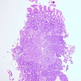
The histological findings shown in figure 1 indicate a proliferation of macrophages and the formation of granulomas in the antrum of the stomach (figure 2). When the presence of granulomas is confirmed in the stomach, a diagnosis of granulomatous gastritis (GG) is made. GG is a type of chronic gastritis and can either be secondary to an underlying condition (further subclassified as infectious or non-infectious) or an isolated finding with no specific identifiable cause (known as idiopathic GG). Idiopathic GG may account for up to 25% of all cases.1

GG is a rare finding, with an incidence of 0.08–0.35%; in the largest series to evaluate its epidemiology 56 cases were identified from a total of 15,947 tissue specimens.2

Patients with GG may exhibit epigastric pain, as in this case, but they may have no symptoms whatsoever. There have also been reports of patients with vomiting and weight loss due to gastric outlet obstruction. Bleeding as a manifestation of GG is exceedingly rare, but it has been seen, especially in patients for whom sarcoidosis is the underlying cause, and it may be massive and life threatening.3 There is also considerable variation in the endoscopic findings, which range from a macroscopically normal mucosa to the appearance of erythema, erosions and gastric rugae hypertrophy. In severe cases, there may be ulcers present or obstruction of the lumen due to mass effect similar to gastric malignancy.

Figure 2 | Histological findings in the stomach of the case patient. a | Figure 1 revisited. Staining of Iron crystalline material within macrophages of the lamina propria of the gastric mucosa (arrows). b | Staining of iron crystalline material within ma

Case Question 2

In view of the past history of this patient, what was the underlying cause of the symptoms observed?

A. Sarcoidosis

B. Granulomatosis with polyangiitis

C. Crohn’s disease

D. Tuberculosis

E. Histoplasmosis

F. A drug-induced reaction

G. Helicobacter pyloriinfection

H. Whipple’s disease

  • Case question 2 answer and discussion
Case question 2 answer and discussion

Correct answer: F.

Discussion

In most patients an underlying cause of GG can be identified. The causes may be classified as infectious or noninfectious. Infectious diseases are the most common aetiology of GG in developing countries. Tuberculosis is an established infectious cause of GG, but it has been also attributed to various other infectious entities, such as Whipple’s disease, syphilis, fungal infections (e.g. cryptococcus, histoplasmosis) and parasitic infections (e.g. taeniasis, anisakiasis).4 An association between GG and H. pyloriinfection has been observed, but in most cases there seems to be another aetiologic agent responsible for the development of the gastritis. Nevertheless, in instances of isolated GG with concurrent H. pylori infection, eradication of H. pylori has led sometimes to improvement.5   

In developed countries, the most frequent causes of GG are noninfectious, with Crohn’s disease and sarcoidosis being the most important. Crohn’s disease is the most commonly encountered underlying condition in patients with GG. Clinically detectable gastritis is found in only 3–6% of patients with Crohn’s disease, but microscopic involvement has been described in up to two thirds of all patients with Crohn’s disease. One third of patients who have microscopic findings may have granulomas. It should also be noted that GG may be encountered as a new finding in patients with known Crohn’s disease, but it can also be the first manifestation of Crohn’s disease especially in children.6,7 Sarcoidosis is another systemic condition that can affect the gastrointestinal tract, with a special preference for the stomach. Microscopic involvement may be seen in 5–10% of patients with sarcoidosis, while only 1% will progress to overt symptoms. Endoscopic findings tend to be more impressive in cases of GG caused by sarcoidosis than in those with other underlying causes, they can range from ulcers to polypoid-like lesions that may cause bleeding or obstruction, respectively.8 Rarer noninfectious causes include vasculitides (e.g. granulomatosis with polyangiitis), tumours (e.g. MALT or adenocarcinoma, especially with a distal oesophageal location) or drugs (e.g. cocaine or interferon) and chronic granulomatous disease in childhood.

A distinct noninfectious aetiological subcategory is GG secondary to foreign body reaction. This is seen as a gastric response to the presence of various substances, such as sutures from previous operations or endogenous materials (e.g. mucin or lipids). A case series has been published that described GG associated with the use of cyanoacrylate glue injections.9 In addition, of particular interest are reports concerning GG presenting after a foreign body reaction to crystalline drugs or agents (e.g. antacids containing aluminium and magnesium). In our case, the histological specimen was highly suggestive for the presence of crystalline material within the macrophages and giant cells. This finding coupled with the patient’s history of taking an iron supplement led to further investigation of the biopsy samples. Staining with Perls’ histochemical stain, more commonly known as Prussian blue, revealed iron crystalline material as the probable cause of the patient’s GG (figure 3).

Figure 3 | Iron crystalline material stained blue. Perls’ histochemical stain (also known as Prussian blue). Magnification x400.

Case Question 2

How should we proceed regarding the management of this patient?

A. Acid suppression with PPIs

B. Investigate other possible causes

C. Stop iron supplementation and take repeat biopsy samples

D. Stop iron supplementation and investigate other possible causes

  • Case question 3 answer and discussion
Case question 3 answer and discussion

Correct answer: D.

Discussion

For our patient, there was a convincing case for GG being caused by iron supplements, but his immunocompromised status warranted further investigation, especially for possible infectious causes. In general, taking a detailed clinical history is essential, as is meticulous histopathological examination with the use of specific stains and/or polarization. Further investigations should then be considered based on the data obtained and pursued according to the degree of clinical suspicion (e.g. ileocolonoscopy for Crohn’s disease, a chest radiograph for tuberculosis or sarcoidosis, measurement of angiotensin-converting enzyme concentrations for sarcoidosis and serum antineutrophil cytoplasmic antibody titres for polyangiitis).

Treatment should be tailored according to the underlying aetiology—in case of a foreign body reaction, removal of the culprit substance is beneficial. Repeat endoscopy is useful for assessing accurately the extent of the condition and at a later time the progression or abatement of granuloma presence.

References

  1. Ectors NL, et al. Granulomatous gastritis: a morphological and diagnostic approach. Histopathology 1993; 23: 55–61. 
  2. Lauwers GY. Gastritis: beyond Helicobacter pylori. Int J Surg Pathol 2010; 18: 28S–32S. 
  3. Akinyemi E, et al. Gastric sarcoidosis. J Natl Med Assoc 2006; 98: 948–949. 
  4. Kamani L, et al. Granulomatous gastritis: a diagnostic dilemma? Singapore Med J 2008; 49: e222–e224. 
  5. Kim YS, et al. A case of H. pylori-associated granulomatous gastritis with hypertrophic gastropathy. Gut Liver 2009; 3: 137–140. 
  6. Laube R, et al. Oral and upper gastrointestinal Crohn's disease.J Gastroenterol Hepatol 2018; 33: 355–364. 
  7. Renault M, et al. Age-related differences in granulomatous gastritis: a retrospective, clinicopathological analysis. J Clin Pathol 2010; 63: 347–350. 
  8. Afshar K, et al. Gastric sarcoidosis and review of the literature. J Natl Med Assoc 2010; 102: 419–422. 
  9. Guner G, et al. Cyanoacrylate associated foreign body granulomatous gastritis: A report of three cases. Case Rep Pathol 2017; 2017: 2753487. 
Social Sharing

Comment

Please log in with your myUEG account to post comments.

Related Articles
Blog
Helicobacter pylori–associated dyspepsia
Decide on the Spot
Which way to go with a giant gastric ulcer?
Decide on the Spot
One finding and two surprises
Mistakes in...
Mistakes in upper gastrointestinal bleeding and how to avoid them
Latest Articles
Decide on the Spot
An elusive lesion in the colon
Decide on the Spot
An incidental diagnosis by endoscopic ultrasound
Decide on the Spot
A not-so-black-and-white case of gastrointestinal bleeding
Decide on the Spot
Yet another case of abdominal pain?
Subscribe
Sign up with your email to receive UEG news and updates. We respect your privacy.

UEG Secretariat

United European Gastroenterology

Wickenburggasse 1
1080 Vienna, Austria


T: +43 1 997 1639
F: +43 1997 1639 10

UEG Week Registration

Mondial Congress & Events

Official UEG Week Registration Partner
Operngasse 20b
1040 Vienna, Austria


T: +43 1 5880 4 0
F: +43 1 5880 4 185

More

Imprint

Terms & Conditions

Media Policies

Privacy Policy

Social Media

16 827 Followers

11 158 Followers

1 784 Followers

1 870 Subscribers

UEG copyright 2021. All rights reserved. Use of this website constitutes acceptance of the Privacy Policy and Terms & Conditions.