• Contact us
  • Young GI
  • Patients
  • Industry
  • Press
Create myUEG account
  • About
    • To landing page
      • Who we are
      • Boards & Committees
      • Governance
      • Secretariat
      • What we do
      • Strategic Drivers
      • Annual Report
      • Our Members
      • Specialist Member Societies
      • National Member Societies
      • Community
      • myUEG
      • Women in GI
      • Young GIs
  • Week
    • To landing page
      • Register
      • Fees
      • CME Accreditation
      • Science
      • Abstracts
      • Programme
      • Postgraduate Teaching
      • Networking
      • Industry Programme
      • Acknowledgements
      •  
      • Congress Resources
      • Week 24/7 recordings
  • Education
    • To landing page
      • Online Education
      • Online Courses
      • Webinars
      • Virtual Summer School
      • Face-to-face Education
      • Masterclass
      • Clinical Visiting Fellowships
      • Education Resources
      • Mistakes in...
      • Articles
      • Image Hub
      • CME in Europe
  • Research
    • To landing page
      • Support
      • Research Prize
      • Research Fellowship
      • Horizon Europe Support
      • Researchers Network
      • Research Switchboard
      • Advocacy
      • Position Papers
      • White Book
  • Quality of Care
    • To landing page
      • Search Guidelines
      • Standards & Guidelines Repository
      • Non-English Guidelines
      •  
      • GI Guidelines App
      • Develop & Implement
      • Essentials for a good guideline
      • Get Funding
      • Guideline Webinars
      • Projects
      • Quality of Care Initiatives
      • Quality Standards
  • Public Affairs
    • To landing page
      • Advocacy
      • Manifesto on Digestive Health
      • Core Positions
      • Position Papers
      • Stakeholder Collaboration
      • MEP Digestive Health Group
      • Key Topics
      • Our Supporters
      • #EUNewsline →
  • Publications
    • To landing page
      • Publications
      • Congress Publications
      • Research Publications
      • Public Affairs Publications
      • Education Publications
      • UEG Publications
      • UEG Journal →
      • UEG Journal Podcast
  • Opportunities
    • To landing page
      • Congress Opportunities
      • National Scholarship Awards
      • Best Abstract Prizes
      • Research Funding
      • Research Prize
      • Research Fellowship
      • Researchers Network
      •  
      • Activities Funding
      • Career Development
      • Rising Star Awards
      • Clinical Visiting Fellowships
      • Open Positions
      • Talent Pool
      • Recognition of Achievements
      • Lifetime Achievement Award
      • Journal Best Paper Award
  • Library
    • To landing page
      • Congress Resources
      • Week 24/7 recordings
      • Abstracts
      • Posters
      • Guideline Resources
      • Education Resources
      • Image Hub
      • Webinars
      • Education articles
  • Contact us
  • Young GI
  • Patients
  • Industry
  • Press
Decide on the Spot

Yet another case of abdominal pain?

May 08, 2018 | Matteo Revelli

A 41-year-old man with a recent history of weight loss, reduced appetite and nausea presents with acute abdominal pain.

On physical examination he has abdominal distension without tenderness but pain during deep palpation, Blumberg’s sign is negative and bowel sounds are sparse. The initial radiography findings are shown in figure 1 and his blood test results are shown in table 1. 

Table 1 | Blood test results at presentation. Click on the picture to enlarge the table.
Figure 1 | Anteroposterior plain radiograph of the abdomen, acquired in standing position, showing air distention of several small bowel loops, associated with the presence of levels suggesting bowel obstruction.

At this stage, would you choose to give laxatives and discharge the patient, urgently perform a CT scan or endoscopy, or give a prokinetic, antibiotics, laxatives and monitor the patient every 6h? The correct decision here is to perform a CT scan, which is what the case patient underwent—the findings are shown in figure 2.

Figure 2 | Abdominal contrast-enhanced CT scan with coronal and sagittal reconstructions; images were acquired at portal venous time (about 70 seconds after injection of iodinated contrast medium).

Case Question 1

What is your clinical diagnosis at this point?

a) Intestinal ischaemia

b) Crohn’s Disease

c) Intussusception

d) Megacolon

  • Case question 1 answer and discussion
Case question 1 answer and discussion

Correct answer: C.

Discussion

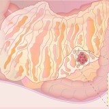
The yellow arrows on figure 3 indicate the presence of small bowel intussusceptions involving different segments of ileal loop in the case patient.

Figure 3 | Small bowel intussusceptions involving different segments of ileal loops are highlighted by the yellow arrows on the multiplanar abdominal contrast-enhanced CT images shown in figure 2.

Intussusception is a serious condition in which part of the intestine slides into an adjacent part of the intestine. This ‘telescoping’ often blocks food or fluid from passing through the intestine. Intussusception also cuts off the blood supply to the part of the intestine that’s affected, which can lead to a tear in the bowel (perforation), infection and death of bowel tissue.1

Intussusception is the most frequent cause of bowel obstruction in paediatric patients;2however, it may occur in adults as the result of an underlying medical condition, such as a tumour, which may act as lead point, dragging part of the bowel into the neighbouring segment.3

There are several medical conditions/situations known to contribute to intussusception in adults:

  • Congenital abnormalities (e.g. Meckel diverticulum, ectopic pancreas, duplication cyst)
  • A polyp or tumour
  • Scar-like tissue (adhesions) in the intestine 
  • Weight-loss surgery (gastric bypass) or other surgery on the gastrointestinal tract
  • Gastrointestinal inflammation caused by disease (e.g. Crohn's disease)

When intussusception occurs in adults, the most frequent symptom is abdominal pain that comes and goes. Nausea and vomiting may also occur. People sometimes have symptoms for weeks before seeking medical attention. A repeat CT scan was performed, and the findings are shown in figure 4.

Figure 4 | Second-look of abdominal contrast-enhanced CT scan with coronal and sagittal reconstructions; images were acquired at portal venous time (about 70 seconds after injection of iodinated contrast medium).

Case Question 2

What is your clinical diagnosis at this point?

a) Congenital abnormality

b) Intestinal lymphoma

c) Polyp syndrome (e.g. FAP, Peutz-Jeghers syndrome, etc.)

d) Metastases

  • Case question 2 answer and discussion
Case question 2 answer and discussion

Correct answer: D.

Discussion

Congenital abnormalities are more likely in young patients and a single area of the bowel is usually involved. 17–30% of small bowel tumours are lymphomas, but they are solitary lesions or involve a single portion of the small bowel (5–20 cm). Furthermore, on imaging they are observed to have the same thickness as the bowel wall. A polyp syndrome is unlikely in this case because FAP involves the colon, while Peutz-Jeghers syndrome involves the small bowel, but melanosis is usually present as well.  Small intestinal metastases are rare and originate mostly from kidney carcinoma or melanoma, but they were suspected to be the cause of the intussusception in the case patient because metastases had been noticed in the liver (figure 5).

Figure 5 | A more accurate evaluation of the images in figure 4 allows the identification of multiple enhancing nodules distributed along the walls of the small bowel, as well of several enlarged lymph nodes within the mesenteric fat (a,b,c). Parenchymal

Biopsy samples were taken from the hepatic nodules and the duodenum was resected, and the findings confirmed metastases of an epithelioid-cell-type melanoma to the enteric wall, liver and peritoneum. The case patient subsequently developed neurological manifestations (figure 6). 

Figure 6 | Unenhanced head CT showing the presence of multiple nodules, involving both cerebral and cerebellar parenchyma, consistent with cerebral metastases.

Metastatic melanoma affects the small bowel more often than the colon.4 Unfortunately, the disease has a late clinical onset and diagnosis is often made only when complications, such as bleeding, obstruction or perforation, occur.  However, there are only few reports of small bowel intussusception caused by melanoma.5 CT is important for diagnosis and staging, followed by histology.6,7 Surgery is often required as is palliative treatment to manage complications (+/- adjuvant chemo/radiotherapy).8

References

  1. Mayo Clinic. Intussusception, www.mayoclinic.org/diseases-conditions/intussusception/.../syc-20351452 (accessed 3 May 2018). 
  2. Bines JE and Ivanoff B. Acute intussusception in infants and children: Incidence, clinical presentation and management: a global perspective. World Health Organization, Department of Vaccines and Biologicals. WHO/V&B/02.19 (2002, accessed 3 May 2018). 
  3. Marsicovetere P, et al. Intestinal Intussusception: Etiology, Diagnosis, and Treatment. Clin Colon Rectal Surg 2017; 30: 30–39. https://www.thieme-connect.com/DOI/DOI?10.1055/s-0036-1593429
  4. Tessier DJ, et al. Melanoma metastatic to the colon: case series and review of the literature with outcome analysis. Dis Colon Rectum2003; 46: 441–447. https://link.springer.com/article/10.1007/s10350-004-6577-2
  5. Schoneveld M, et al.Intussusception of the small intestine caused by a primary melanoma? Case Rep Gastroenterol 2012; 6: 15–19. https://www.ncbi.nlm.nih.gov/pmc/articles/PMC3290021/
  6. Li Q and Yang J. Role of imaging methods in metastatic melanoma. Onkologie2012; 35: 522–526. https://www.karger.com/Article/Abstract/341833
  7. de Waal AC, et al. Melanoma of unknown primary origin: a population-based study in the Netherlands. Eur J Cancer 2013; 49: 676–683. https://www.ejcancer.com/article/S0959-8049(12)00697-1/fulltext
  8. Kamposioras K, et al. Malignant melanoma of unknown primary site. To make the long story short. A systematic review of the literature. Crit Rev Oncol Hematol2011; 78: 112–126. https://www.croh-online.com/article/S1040-8428(10)00092-2/fulltext
Social Sharing
  • About the Author
About the Author

Matteo Revelli works as consultant at Reggio Emilia AUSL - IRCCS Department of Diagnostic Imaging and Laboratory Medicine in Reggio Emilia, Italy. He graduated in medicine in 2008 and undertook his radiology postgraduate education in 2014 at Genoa University. His main clinical and research fields of interest include gastroenterological, cardiac and urogenital radiology. He is author or coauthor of more than 80 papers, including scientific articles indexed on Pubmed, multimedia publications, posters and oral presentations presented at national and international conferences

Comment

Please log in with your myUEG account to post comments.

Related Articles
Mistakes in...
Mistakes in microscopic colitis and how to avoid them
Mistakes in...
Mistakes in endoscopic ultrasonography and how to avoid them
Mistakes in...
Mistakes in nutrition in IBD and how to avoid them
Press Release
Turning the tide on Europe’s deadliest cancer: tackling the burden of pancreatic cancer
Latest Articles
Decide on the Spot
An elusive lesion in the colon
Decide on the Spot
A rare biopsy finding with many possible causes
Decide on the Spot
An incidental diagnosis by endoscopic ultrasound
Decide on the Spot
A not-so-black-and-white case of gastrointestinal bleeding
Subscribe
Sign up with your email to receive UEG news and updates. We respect your privacy.

UEG Secretariat

United European Gastroenterology

Wickenburggasse 1
1080 Vienna, Austria


T: +43 1 997 1639
F: +43 1997 1639 10

UEG Week Registration

Mondial Congress & Events

Official UEG Week Registration Partner
Operngasse 20b
1040 Vienna, Austria


T: +43 1 5880 4 0
F: +43 1 5880 4 185

More

Imprint

Terms & Conditions

Media Policies

Privacy Policy

Social Media

16 827 Followers

11 158 Followers

1 784 Followers

1 870 Subscribers

UEG copyright 2021. All rights reserved. Use of this website constitutes acceptance of the Privacy Policy and Terms & Conditions.