Frans JC Cuperus is a resident in the Department of Gastroenterology and Hepatology of the Radboudumc, Nijmegen, The Netherlands. His research interests include hereditary hyperbilirubinaemias and canalicular transporter defects in cholestasis.
Professor Dr Joost PH Drenth is a hepatologist and the current chair of the Department of Gastroenterology and Hepatology of the Radboudumc, Nijmegen, The Netherlands. His main research interests include polycystic liver disease, autoimmune hepatitis and hepatitis C.
Eric T Tjwa is a hepatologist in the Department of Gastroenterology and Hepatology of the Radboudumc, Nijmegen, The Netherlands. His research interests include fibrogenesis of the liver, viral hepatitis and complications of liver cirrhosis, including hepatocellular carcinoma.

Liver function tests (LFTs) are routinely used to screen for liver disease. A correct interpretation of LFT abnormalities may suggest the cause, severity, and prognosis of an underlying disease. Once the diagnosis has been established, sequential LFT assessment can be used to assess treatment efficacy.
Abnormal LFTs are frequently encountered in clinical practice, since elevation of at least one LFT occurs in more than 20% of the population.1 Many patients with abnormal LFTs, however, do not suffer from structural liver disease, since these tests can be influenced by factors unrelated to significant liver damage or liver function loss. During normal pregnancy, for example, serum albumin levels fall due to plasma volume expansion, and alkaline phosphatase (ALP) levels rise due to placental influx. Patients who have elevated transaminase levels may not suffer from liver disease, but rather from cardiac or skeletal muscle damage. Conversely, patients who suffer from advanced liver disease, such as chronic hepatitis or compensated liver cirrhosis, may have normal LFTs.
In short, the assessment of LFTs can represent a challenge for physicians. The observations above demonstrate the need for a firm understanding of the individual LFT, and the ability to interpret the results in the light of a specific clinical setting. Such an understanding is not merely a goal on its own, but may serve as a template to avoid mistakes in interpreting LFT abnormalities.
In the following sections, we discuss several mistakes frequently made in the interpretation of LFTs and how to avoid them. Most of the discussion is evidence based, but where evidence is lacking the discussion is based on extensive clinical experience.
© UEG 2017 Cuperus, Drenth and Tjwa.
Cite this article as:
Cuperus FJC, Drenth JPH and Tjwa ET. Mistakes in liver function test abnormalities and how to avoid them. UEG Education 2017: 17; 1–5.
Correspondence to:
Conflicts of interest:
The authors declare there are no conflicts of interest.
Published online:
January 26, 2017.
FThe term liver function test is a misnomer, since most LFTs do not measure the function of the liver, but are markers of liver injury. Indeed, most LFTs should actually be referred to as liver tests.
Alanine aminotransferase (ALT) and aspartate aminotransferase (AST) are biochemical markers of hepatocellular injury. During hepatocellular injury, these enzymes leak into the systemic circulation. ALT is localized in the cytosol and AST both in the cytosol and mitochondria of hepatocytes.2 AST can also be found in other tissues, such as skeletal and cardiac muscle and red blood cells. Of note, the upper limit of normal (ULN) cut-off value for serum ALT levels is slightly higher in men than in women.3
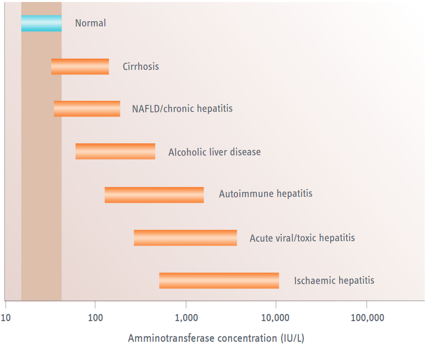
The magnitude of the aminotransferase elevation provides important clues regarding the cause of the hepatocellular injury (figure 1).
- Patients with compensated cirrhosis, chronic viral hepatitis (B or C) or nonalcoholic fatty liver disease (NAFLD) have normal or mildly elevated AST and ALT levels.4,5 Levels of >5 x ULN indicate concomitant acute liver injury.
- Alcoholic liver disease (ALD) is associated with AST levels of <8 x ULN and ALT levels of <5 x ULN.6
- Acute viral and toxic hepatitis with jaundice are associated with AST and ALT levels of >25 x ULN.7,8
- AST and ALT levels are highest (up to >50 x ULN) during ischaemic liver injury (shock liver, ischaemic hepatitis).9
In addition, the AST:ALT ratio can be used to interpret the underlying cause of the aminotransferase elevation. An AST:ALT ratio of ≥2:1 is suggestive (≥3:1 highly suggestive) of ALD.6,10,11 The relatively low ALT level in patients with ALD is caused by depletion of pyridoxine (vitamin B6), which is used as a coenzyme in the synthesis of both AST and ALT.12 ALT synthesis, however, is more affected than AST synthesis. Alcohol also induces mitochondrial injury, which releases mitochondrial AST. Mitochondrial AST has a longer half-life compared with ALT or cytosolic AST (~87 h versus ~47 h and ~17 h, respectively) and can thus be detected for a longer period of time after cessation of alcohol intake.9 With abstinence from alcohol, an ALT:AST inversion generally occurs within 30–90 days in the absence of significant concomitant liver disease.

Figure 1 | Elevation of aminotransferase concentrations in hepatocellular injury. NAFLD, nonalcoholic fatty liver disease.
Gamma glutamyl transferase (GGT) is a highly sensitive, but nonspecific enzyme marker for liver disease. GGT is expressed in the epithelial cell membranes of various tissues.13 In the liver, GGT is mainly expressed in biliary epithelial cells. The GGT test is mainly useful in two situations:
- An elevated level of GGT in the presence of an AST:ALT ratio of >2:1 suggests alcohol-related liver disease, and can be used to monitor alcohol abstinence (GGT levels return to normal after 2–6 weeks of abstinence).6,14
- Unlike ALP levels, GGT levels do not rise during bone disease. A simultaneous increase in ALP and GGT thus confers liver specificity to serum ALP elevation.13
Although an isolated increase in the GGT conce
ntration lacks specificity for detecting liver disease or alcohol abuse, its diagnostic merit lies in having an excellent negative predictive value (NPV) for hepatobiliary disease.1 The serum GGT level is rarely normal during intrahepatic cholestasis. The exception to this rule occurs in patients who have familial intrahepatic cholestasis (PFIC) type 1 and 2, since these patients suffer from severe inherited cholestatic liver disease in the presence of normal GGT levels. By contrast, patients with PFIC type 3 usually have a milder phenotype, but a marked isolated elevation in their GGT levels.15
ALP is a nonspecific marker of liver disease that is mainly expressed in the liver, but also in bone, intestine and placenta. Liver and bone disease are the most frequent causes of pathological elevation of ALP levels. Isolated elevation of ALP levels (e.g. without elevation of GGT levels) may necessitate ALP fractionation in order to determine its hepatic origin.16 The ALP test is useful for detecting intrahepatic and extrahepatic biliary obstruction, but is less sensitive than GGT.
- Intrahepatic and extrahepatic biliary obstruction is usually associated with an ALP level of >4 x ULN. This elevation is due to an increase in ALP synthesis, and may take 1–2 days to develop. After resolution of the obstruction, normalization of ALP levels may take several days because its half-life is 7 days.9
- Persistently elevated ALP levels in the absence of biliary obstruction warrant determination of anti-mitochondrial antibodies (AMA), which are highly specific for primary biliary cholangitis (PBC).17 In PBC patients, ALP levels can be used to monitor treatment response and are associated with transplant-free survival.18
Bilirubin is produced in the mononuclear phagocyte system from the breakdown of haem, which is mainly derived from senescent red blood cells.19 After formation, most bilirubin is reversibly bound to plasma albumin and transported to the liver. Once in the liver, bilirubin is conjugated by the enzyme UDP-glucuronosyltransferase, which renders the molecule more water soluble and thereby allows its excretion into the bile.20 In the intestine, conjugated bilirubin is hydrolysed to unconjugated bilirubin by the enzyme β-glucuronidase and broken down by the intestinal microflora to urobilinogen and other urobilinoids. These urobilinoids are partly reabsorbed and can spill over from the enterohepatic circulation into the systemic bloodstream.21 Normal urine contains urobilinogen, but conjugated bilirubin only spills into the urine during conjugated hyperbilirubinaemia.
- Isolated unconjugated or conjugated hyperbilirubinaemia does not usually reflect significant cholestatic or hepatocellular liver damage.
- Conjugated hyperbilirubinaemia in the presence of other LFT abnormalities may be due to either extrahepatic cholestasis, hepatocellular damage or infiltrative liver disease. Plasma conjugated bilirubin levels thus have no meaningful role in differentiating between these conditions.
- Conjugated bilirubin levels are a marker for the excretory capacity of the liver, and can be used to predict prognosis in advanced liver disease. Consequently, plasma bilirubin levels are a component of the Model for End Stage Liver Disease (MELD) and Child–Pugh scores.22,23 The MELD score is used to determine prognosis and improve the organ allocation system for liver transplantation, whereas the Child–Pugh score predicts 1-year and 2-year survival in advanced chronic liver disease.
Albumin is the most abundant plasma protein, and is responsible for 75% of the plasma colloid osmotic pressure.24 Albumin synthesis occurs exclusively in the liver, and can be doubled during albumin loss or dilution. Albumin has a long half-life (14–20 days), and the plasma albumin concentration is consequently not useful as a marker of liver synthesis during acute liver disease.9 By contrast, plasma albumin is an excellent marker of liver function during advanced chronic liver disease (e.g. cirrhosis) and is thus a component of the Child–Pugh score.
The prothrombin time measures the time taken for prothrombin to be converted to thrombin via the extrinsic coagulation pathway. This pathway depends on the coagulation factors II, V, VII, and X, which are all produced in the liver. Factor VII has a plasma half-life of only 6 hours, and the prothrombin time is consequently useful as a marker of liver synthesis during acute liver disease. Massive hepatocellular necrosis (>80%) during toxic hepatitis, for instance, can lead to an increased prothrombin time in the presence of normal plasma albumin levels. Conversely, the prothrombin time may remain completely normal during compensated cirrhosis until a marked decrease in liver function occurs. The prothrombin time is not a reliable marker of bleeding risk in patients who have cirrhosis, since it does not take the production of anticoagulant factors (e.g. protein C, protein S) into account, which is reduced in these patients.25
Differences in laboratory assays mean that prothrombin time is currently reported as the international normalized ratio (INR), which allows for standardization across laboratories. Prothrombin time is a component of both the MELD and Child–Pugh score. In addition, factor V activity is integrated in the Clichy score, which is used to predict mortality and evaluate the necessity for liver transplantation during acute liver failure (table 1).26
| Scoring system | Prognostic factors |
| King’s College criteria for OLT |
Acetaminophen-induced ALF
Non-acetaminophen-induced ALF Encephalopathy present (irrespective of grade) and:
|
| Clichy criteria for OLT | Presence of hepatic encephalopathy and:
|
Table 1 | Scoring systems for severity of acute liver failure/necessity of transplantation. ALDF, acute liver failure; INR, international normalozied ratio; OTL, orthotopic liver transplantation.
None of the LFTs discussed in this article is 100% liver specific. The possibility of a nonhepatic origin of LFT abnormalities should, therefore, always be considered. This holds especially true for isolated LFT abnormalities.
ALT is more liver specific than AST, since the latter can also be found in skeletal and cardiac muscle, kidneys, brain, lungs, pancreas and red blood cells.3 A disproportionate or isolated AST elevation, therefore, should raise suspicion that the source is nonhepatic. Nonhepatic causes of AST elevation include injury to skeletal or cardiac muscle, hyperthyroidism or hypothyroidism, haemolysis, and (rarely) macro-aspartate aminotransferase. The latter condition is caused by the binding of AST to immunoglobulins, which results in delayed AST clearance.27
GGT is expressed in the kidney, pancreas, spleen, lung, heart and brain.13 In general, an isolated elevation of GGT levels is not a specific marker for liver disease, since it can be elevated in patients with diabetes, chronic obstructive pulmonary disease, myocardial infarction, pancreatic disease or renal failure. GGT levels can also be elevated in patients using enzyme inducers (CYP2C, CYP3A, CYP1A) such as phenobarbital, carbamazepine or alcohol.28
ALP consists of several isoenzymes that are located in liver (isoenzyme 1 and 2), bone, intestine and placenta. ALP can be fractionated in order to determine its origin. Bone-derived ALP is increased in patients who suffer from bone disease (e.g. Paget’s disease, primary and metastatic bone tumours, osteomalacia, rickets, hyperparathyroidism), and in children due to rapid bone growth. Intestinally-derived ALP is increased in patients with blood group O or B after fatty meals, and in those with familial ALP elevation.9 Raised intestinal ALP isoenzyme levels have also been reported in patients with liver cirrhosis, diabetes, chronic kidney disease, and bowel ischaemia.30 The placental ALP isoenzyme can be elevated in pregnant women, usually during the third trimester.9 The Regan isoenzyme, a rare variant of placental ALP, can be elevated in cancers that do not involve the bone, such as gonadal, urologic or lung cancer.31 Of note, after the age of 50 years, ALP levels (both hepatic and bone) tend to increase, especially in women.29
Hypoalbuminaemia can have various nonhepatic causes, such as a decrease in albumin synthesis (e.g. malnutrition, malabsorption), albumin dilution (e.g. pregnancy), albumin loss (e.g. nephrotic syndrome, protein-losing enteropathy), or a catabolic state (e.g. infection, trauma, malignancy). Hypoalbuminaemia without liver test abnormalities is usually not associated with liver disease.
The prothrombin time can be affected by various coagulation disorders in the absence of hepatic disease, such as disseminated intravasal coagulation and conditions that affect the function of vitamin K (which activates clotting factors II, VII and X of the intrinsic coagulation pathway). These conditions include the use of warfarin and vitamin K deficiency during cholestatic liver disease and cirrhosis, which occurs due to a decrease in its intestinal absorption.32
Acute liver failure is characterized by the development (in days to weeks) of acute massive liver injury (e.g. aminotransferase elevation), jaundice, coagulopathy (INR >1.5), and encephalopathy in the absence of chronic liver disease.33 The condition usually carries a very poor prognosis unless orthotopic liver transplantation is performed. A correct diagnosis and the ability to predict the need for liver transplantation are thus of the utmost importance.34 Several scoring systems, such as the King’s College and Clichy criteria, have been proposed to assess the necessity for liver transplantation.26,35
The significance of aminotransferase levels in the diagnosis and prognosis of acute liver failure is often misunderstood. Excessive aminotransferase levels occur in acute viral, toxic or ischaemic liver injury. Although impressive, these levels merely reflect acute hepatocellular damage rather than loss of liver function. Consequently, marked aminotransferase elevations in the absence of jaundice, coagulopathy and encephalopathy should not lead to a diagnosis of acute liver failure.33
In addition, plasma aminotransferase levels are often markedly elevated at the onset of acute liver failure, and accompanied by relatively modest elevations in bilirubin and ALP levels. If hepatic failure progresses, the hepatocellular pattern usually becomes mixed or even cholestatic as the aminotransferase levels fall. Although a decrease in aminotransferase levels can indicate spontaneous recovery, it may represent worsening of liver failure due to a decrease in hepatocellular mass. Such progression of hepatic failure is typically accompanied by a rise in bilirubin levels and INR, and carries a poor prognosis.23 Conversely, a decrease in aminotransferase levels accompanied by bilirubin and INR normalization indicates recovery of liver failure.
Excessive alcohol consumption is associated with a wide range of hepatic manifestations, including hepatic steatosis, steatohepatitis, fibrosis, cirrhosis and hepatocellular carcinoma. Alcoholic hepatitis and cirrhosis are both associated with significant morbidity and mortality in the setting of continued alcohol abuse. A reliable history is pivotal in establishing the diagnosis, but this may not always be forthcoming. Marked elevations in aminotransferase levels (>8 x ULN) are atypical for alcoholic liver disease and should raise suspicion of concurrent (e.g. ischaemic, toxic or viral) liver injury.6 The most frequent laboratory abnormality in alcoholic hepatitis is an increase in the plasma bilirubin level, whereas aminotransferase levels usually remain below 300 U/L, and rarely rise beyond 500 U/L.36 Other LFT abnormalities in alcoholic hepatitis include an AST:ALT ratio of >2:1 in the presence of elevated GGT levels, and an increase in prothrombin time.
JALT and AST levels are excellent markers for acute choledocholithiasis, since their elevation is usually the first laboratory abnormality that occurs following acute biliary obstruction. Only later will increases in plasma bilirubin, ALP and GGT eclipse ALT and AST levels. In addition, increased ALT levels (>150 IU/L) have a positive predictive value (PPV) of 95% for a biliary aetiology of acute pancreatitis.37
Isolated hyperbilirubinaemia can be predominantly unconjugated (>80% of total) or conjugated (>50%), and does normally not reflect significant liver disease. Isolated unconjugated hyperbilirubinaemia is usually caused by Gilbert’s syndrome, an inherited defect in bilirubin conjugation caused by polymorphisms in the promotor of the UDP-glucuronosyltransferase gene that occurs in ~10% of the general population.38 Patients with Gilbert’s syndrome suffer from a mild, recurrent, unconjugated hyperbilirubinaemia that is exacerbated after fasting, strenuous exercise or intercurrent illness.39 Therapy is not required, and the most important aspect of care involves recognition of the disorder and its benign nature.40
Unconjugated hyperbilirubinaemia may also occur secondary to haemolytic disease, which results in excessive bilirubin production. Unconjugated bilirubin levels in haemolytic anaemia do usually not exceed 80 µmol/L, but may increase further in the presence of Gilbert’s syndrome.41 Isolated conjugated hyperbilirubinaemia occurs in individuals with Rotor or Dubin–Johnson syndrome, both of which are rare and usually manifest during childhood. These syndromes are caused by genetic defects in the hepatic uptake/storage (Rotor) and excretion (Dubin–Johnson) of conjugated bilirubin.42
Drug-induced liver injury (DILI) refers to liver injury caused by drugs, phytotherapeutics, and other potentially toxic substances. DILI can mimic almost every clinical pattern of liver disease, and the identification of an offending agent can be challenging. The diagnosis of DILI is based on three criteria: (1) a temporal (chronologic) relationship with the offending drug, (2) exclusion of other possible causes, and (3) knowledge of the drug’s hepatotoxic potential and its signature pattern. A detailed history is key in identifying a temporal relationship between recently used drugs and the onset of symptoms. This history should include prescription medications, over-the-counter preparations, vitamins, dietary supplements and herbals. As an example, in patients who have AST and ALT levels of >25 x ULN, a detailed history of acetaminophen use is essential. The website livertox.nih.gov provides essential information regarding the hepatotoxic potential and signature pattern of drugs, and should consequently be consulted if DILI is suspected.
-
About the Authors
-
Your liver function test abnormalities briefing
UEG Week
- ‘IBD and abnormal liver tests’ presentation at UEG Week 2016.
- ‘Deranged liver and pancreatic biochemistry: What to do?’ session at UEG Week 2014.
- ‘When do we need to assess liver function?’ session at UEG Week 2014.
- ‘Common presentations in liver disease: Abnormal liver tests’ presentation at UEG Week 2013.
-
‘Common presentations in liver disease: Abnormal liver tests’ syllabus contribution at UEG Week 2013.
Society conferences - ‘Common LFTs in pediatric hepatology’ presentation as ESPGHAN Pediatric Hepatology Summer School 2014.
- ‘Investigation of patients with raised transaminases’ presentation, questions and discussion in the Hepatology session at ASNEMGE 2012.
Standards and Guidelines
- Kwo PY, Cohen SM and Lim JK. ACG Clinical Guideline: Evaluation of abnormal liver chemistries. Am J Gastroenterol 2017; 112: 18–35.
- European Association for the Study of the Liver and Asociación Latinoamericana para el Estudio del Hígado. EASL-ALEH Clinical Practice Guidelines: Non-invasive tests for evaluation of liver disease severity and prognosis. J Hepatol 2015; 63: 237–264.
Please log in with your myUEG account to post comments.